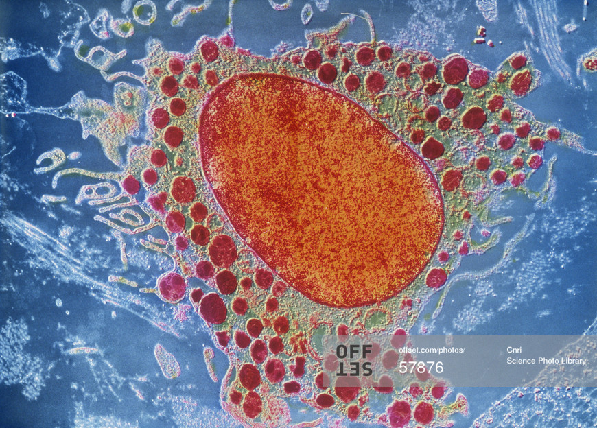
Mast cell, coloured transmission electron micrograph.
Investigating roles of IL-10 as a co-stimulator of mast cell-mediated allergic responses.
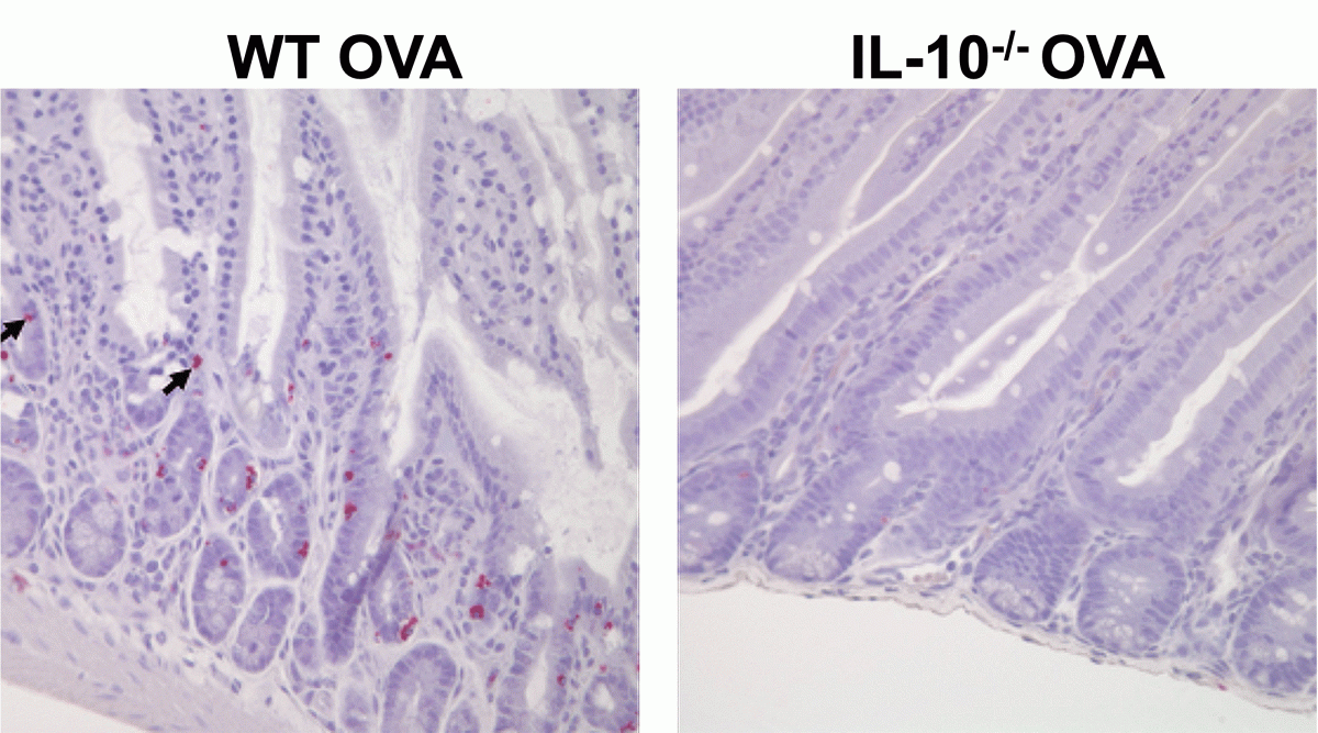
We have previously demonstrated that IL-10 can act as a potent stimulator of mast cells during allergic responses. IL-10 not only enhanced IgE-mediated mast cell activation and function, but also promoted mast cell homeostasis and expansion during food allergy development. Furthermore, the induction of food allergy was attenuated in IL-10-/- mice, which corresponded with decreased systemic mast cell activation and fewer intestinal mast cells. This could be corrected through the transfer of either wild-type mast cells or CD4 T cells, suggesting that the inhibition of food allergy in IL-10-/- mice is due to a lack of IL-10-responsive mast cells and CD4 T cell-derived IL-10 is sufficient to restore this.
In this project, funded by the NIH/NIAID, we further characterize the effects of IL-10 on mast cells, with the goal of elucidating whether IL-10’s effects during food allergy are conferred on mucosal mast cells (MMCs), connective tissue mast cells (CTMCs), or both. Furthermore, we elucidate whether both autocrine and paracrine IL-10 are involved in the regulation of mast cell homeostasis and function. Finally, we investigate whether the “source” of IL-10 and “the inflammatory context” make a difference: i.e., are IL-10’s effects different depending on which cell produces it?
Allergen exposure induces the recruitment of mast cells to the intestines of wild-type but not IL-10-deficient mice.
Assessing mechanisms underlying the IL-33-mast cell axis during allergic responses
While historically, the IgE-mediated activation of mast cells, has been thought to be the primary culprit in allergic diseases, several recent lines of evidence indicate that non-IgE-mediated mast cell activation can also play critical roles during allergic inflammation. Of these, the role of IL-33-responding mast cells is becoming increasingly important.
Current projects have been investigating several variables with the potential to modulate IL-33-mediated mast cell function. These include the role of IL-10 as described above, dietary factors, and epigenetic modifications.
Role of epigenetic modifications in regulating mast cell responses
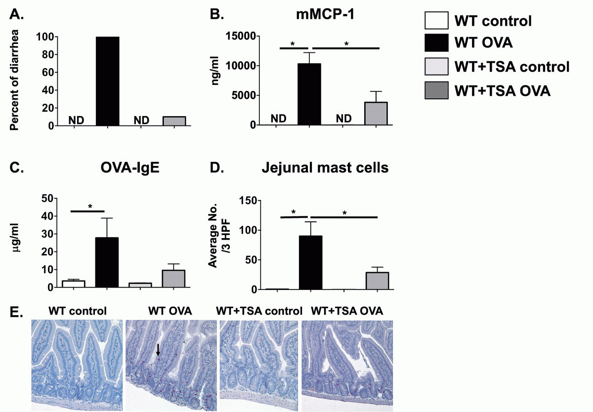
Epigenetic modifications such as histone acetylation and DNA methylation can play a major role in cellular activation and function by regulating the magnitude of gene expression. We have found that modulation of histone acetylation in mast cells by blocking the activity of histone deacetylases can inhibit IgE and IL-33-mediated mast cell function, which extended to suppression of mast cell-mediated responses during food allergy in vivo. Current projects in the lab aim at examining the mechanisms underlying the regulation of mast cell responses through epigenetic modification.
Modulation of Mast cell-mediated responses during Food Allergy by Trichostatin A (TSA)
Regulation of mast cell-mediated responses by dietary factors
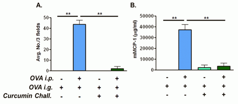
Our lab is very interested in the mechanisms by which dietary factors modulate mast cell activation and the development of food allergy. We have previously shown that curcumin, the active ingredient of the curry spice turmeric, can suppress mast cell responses during food allergy, leading to protective effects. Current projects evaluate the role of curcumin and other dietary factors in modulating mast cell responses and ascertaining the mechanisms involved.
Curcumin ingestion during OVA challenge inhibits intestinal mast cell numbers (A) and mast cell activation (B).
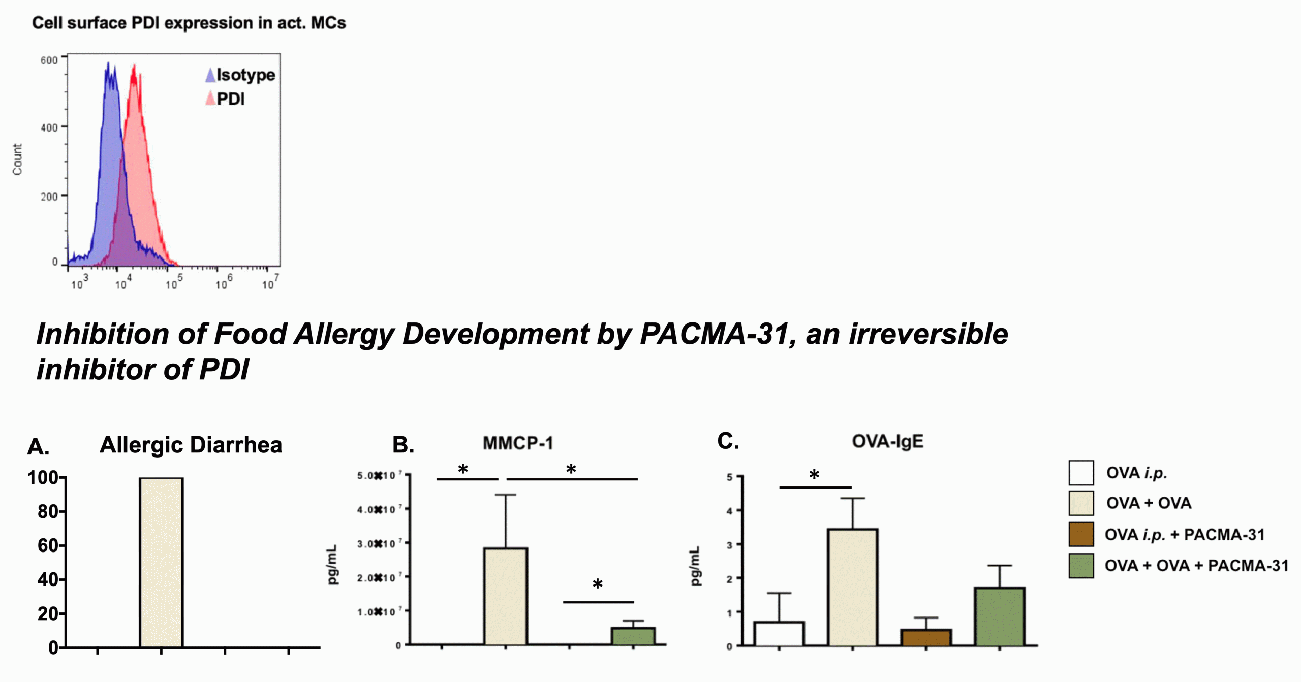
Examination of the role of protein disulfide isomerases (PDIs) in mast cell activation
PDIs are important housekeeping molecular chaperones that play critical roles in cellular function and survival. PDIs help catalyze the formation of disulfide bridges in proteins, thus playing an important role in protein folding and consequently its function. We found that mast cells can express PDI on the cell membrane and that this extracellular PDI can contribute to mast cell activation and function. Blocking PDI using natural and synthetic inhibitors suppressed mast cell activity and attenuated the development of food allergy in mice. Current projects are aimed at investigating the mechanisms by which mast cell-specific PDI can regulate its function.